Mole Removal
Non-Surgical Mole Removal with Laser & Radiofrequency
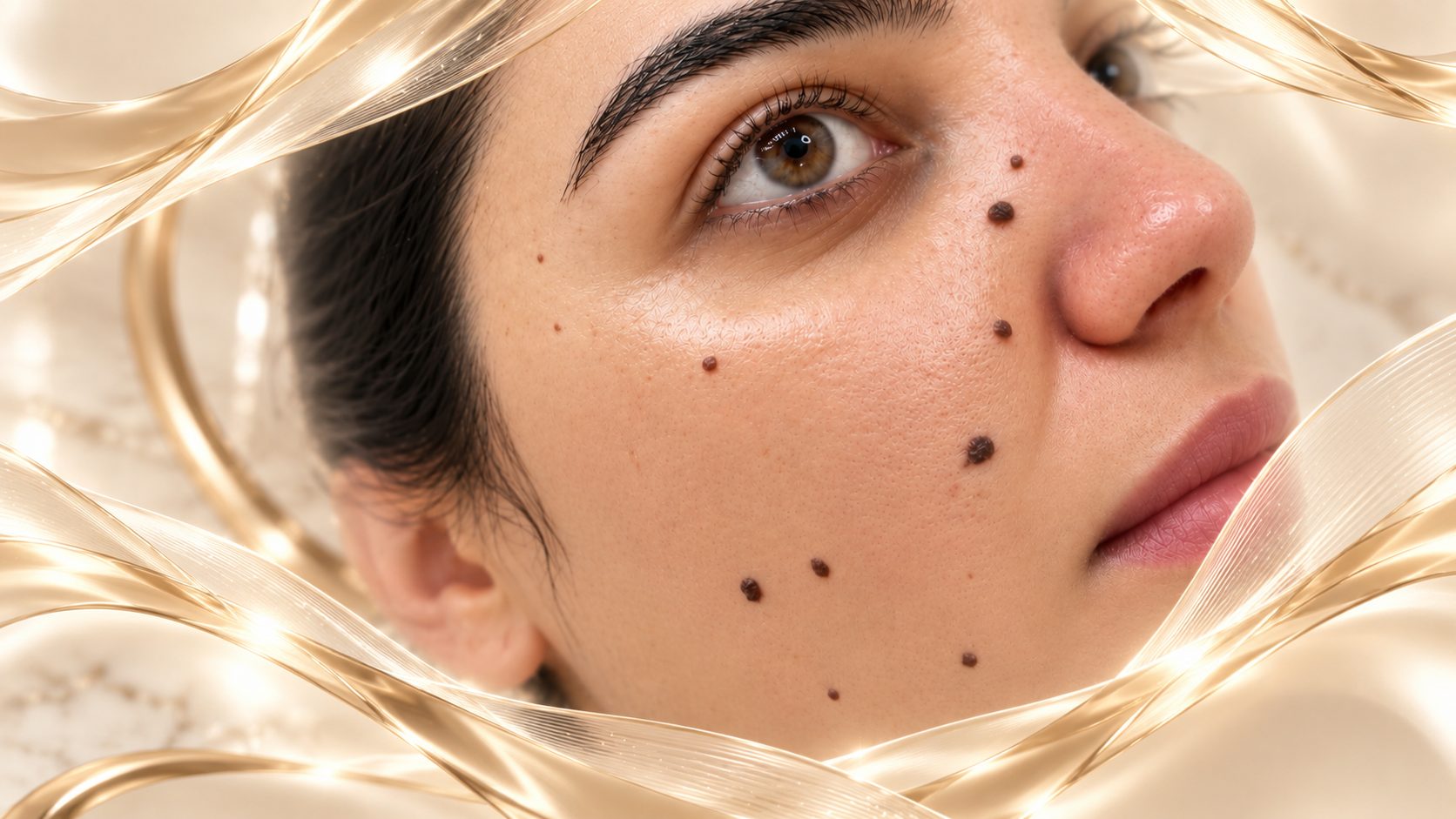
Most moles are benign, but any lesion that is changing, unusual or clinically suspicious should be assessed before cosmetic treatment.
At Dr Ahmet Demir Clinic, suitable benign lesions may be treated with laser or radiofrequency after clinical and, when appropriate, dermoscopic assessment. The priority is medical safety first, followed by the best possible cosmetic outcome.
Contents
What Is Non-Surgical Mole Removal?
For selected benign raised or superficial lesions, laser or radiofrequency energy can be used to remove or reduce the visible lesion without a conventional surgical incision.
Radiofrequency uses controlled thermal energy, while laser treatment uses targeted light energy. The method is selected according to the lesion's appearance, depth and location.
Which Types of Lesions May Be Suitable?
Selected benign facial or neck moles, small raised lesions, skin tags and some superficial pigmented lesions may be considered after examination.
Red vascular lesions such as cherry angiomas require a different treatment approach from melanocytic moles.
Lesions around the eyelids, beard, scalp or other delicate areas require particularly careful assessment.
Dermoscopic Assessment and Safety
Before cosmetic removal, the lesion is assessed for features such as asymmetry, border irregularity, colour variation and clinical change.
If a lesion appears atypical or suspicious, cosmetic ablation is not the appropriate first step. Surgical assessment and histopathological examination may be recommended instead.
Will There Be a Scar?
No procedure can guarantee completely scar-free healing. With appropriately selected superficial benign lesions, non-surgical techniques can often avoid a linear surgical scar, but temporary redness, pigment change or a small residual mark may occur.
Healing depends on lesion depth, location, skin type and aftercare.
How Long Does the Procedure Take?
Small lesions can often be treated in a short appointment under local anaesthesia or topical anaesthetic when appropriate.
Multiple lesions may require a longer session or staged treatment.
Aftercare
A small crust or superficial healing area may form after treatment.
Do not pick at the treated area. Follow the recommended wound-care and sun-protection instructions to reduce the risk of pigment change.
When Should a Mole Be Checked Promptly?
Seek medical assessment for a mole or skin lesion that changes in size, shape or colour, bleeds without clear cause, repeatedly ulcerates, becomes persistently itchy or looks distinctly different from your other moles.
Our Approach
We do not treat every mole cosmetically simply because a patient wants it removed. The lesion must first be considered appropriate for the selected method.
Message us for a free preliminary assessment
Send your photo via WhatsApp and we'll get back to you with a physician's assessment on suitability and planning.